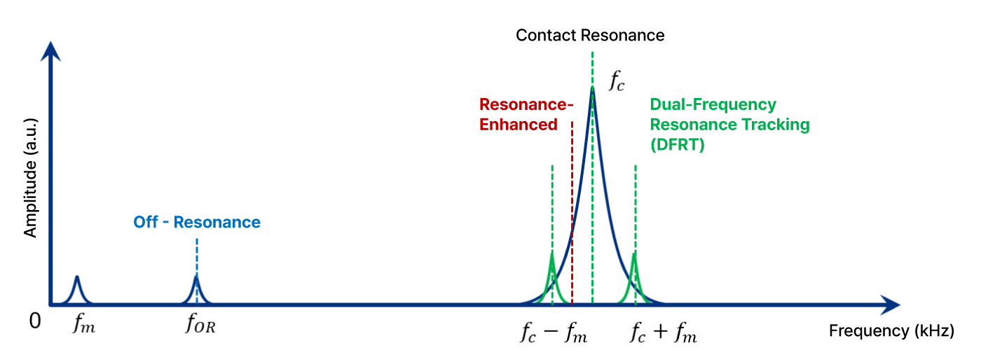
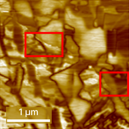
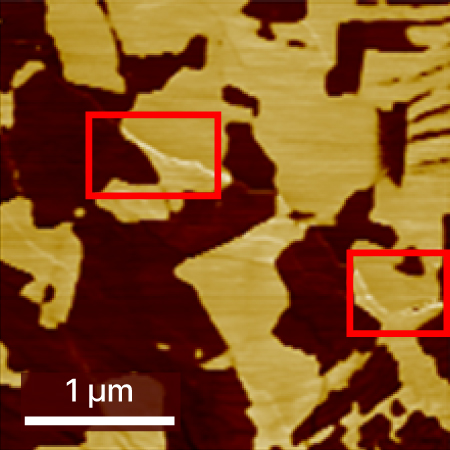
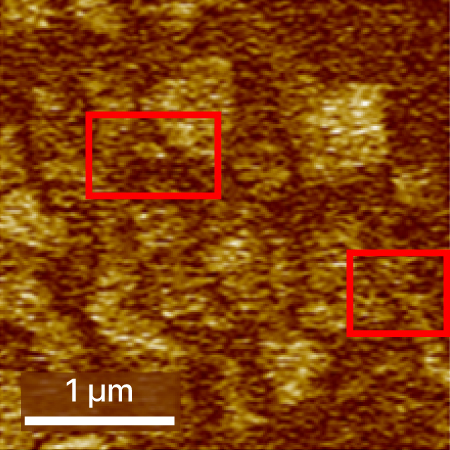
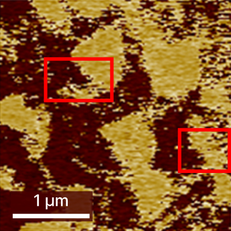
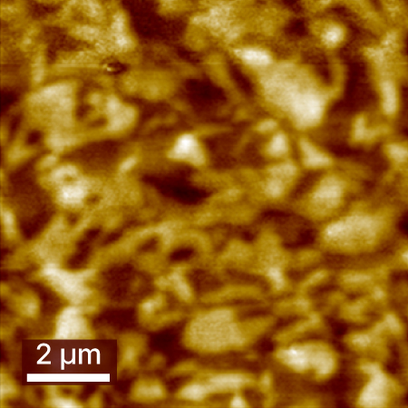
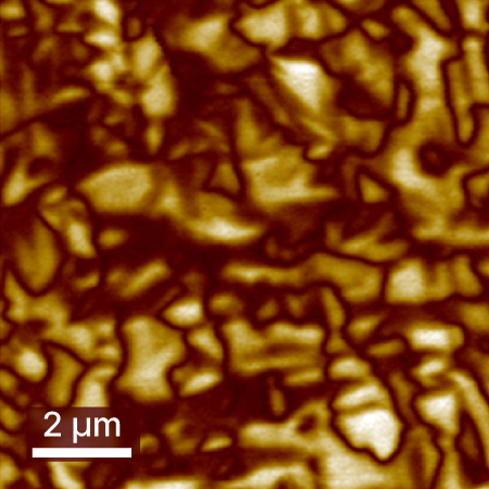
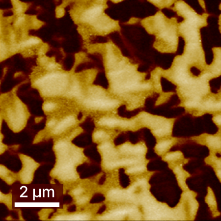
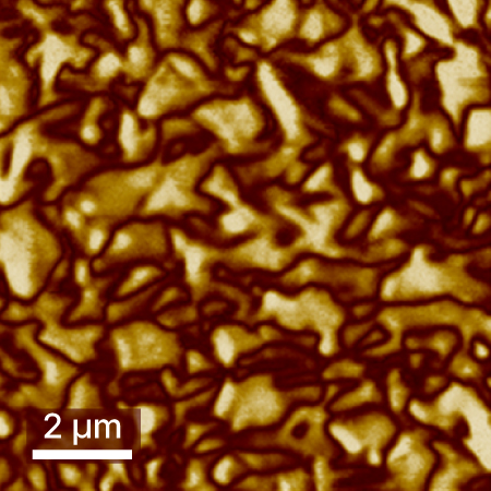
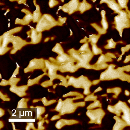
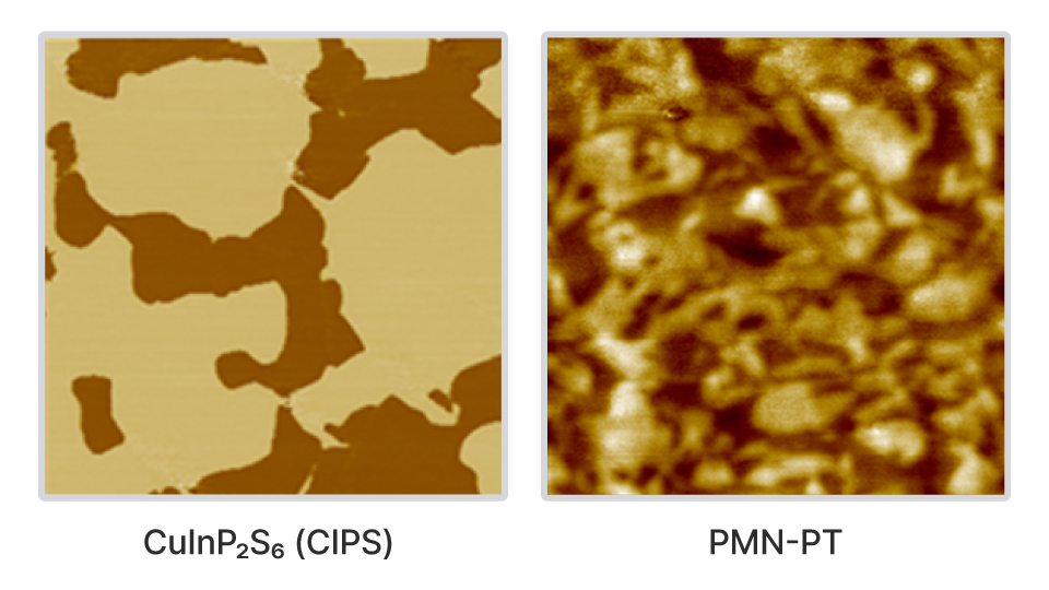
Dual Frequency Resonance Tracking-Piezoresponse Force Microscopy (DFRT-PFM)
High-sensitivity piezoresponse imaging using dual-frequency resonance tracking for precise analysis of complex domain structures and electromechanical behavior
What is DFRT-PFM
DFRT-PFM is an advanced piezoresponse imaging technique developed by Park Systems to overcome the limitations of conventional PFM. By using two frequencies for resonance tracking and another for actual piezoresponse detection, DFRT-PFM maintains stable, high-sensitivity measurements even as the resonance conditions change. This enables precise visualization and analysis of complex domain structures, polarization switching, and local electromechanical responses in a wide range of functional materials.
Reasons to Use This Mode
For a technical comparison between DFRT PFM and conventional PFM techniques, like Off resonance-PFM (OR-PFM) and Contact resonance-PFM (CR-PFM), measurements were carried out on the same region of the CuInP₂S₆ (CIPS) sample using consistent experimental parameters. The collected datasets consist of surface topography (height channel) along with vertical PFM amplitude and phase images from each method. DFRT PFM provides high-resolution feature delineation with minimal lateral noise. Conversely, conventional techniques display lateral noise artifacts, and OR-PFM notably fails to resolve many features. In the boxed regions of the images, a diagonal boundary—present in both topography and CR-PFM—is nearly indistinguishable in OR-PFM data. Such artifacts complicate accurate differentiation of true piezoresponse signals from topographic crosstalk, especially when the domain structure is unknown, potentially compromising data interpretation.
Height
  • Sample: CuInP₂S₆ (CIPS)
  • System: FX40
  • Scan Size: 3 µm × 3 µm
DFRT-PFM Amplitude
DFRT-PFM Phase
CR-PFM Amplitude
CR-PFM Phase
OR-PFM Amplitude
OR-PFM Phase
Applications and Use Cases
A PMN-PT single crystal was measured using DFRT-PFM to demonstrate simultaneous vertical and lateral piezoresponse imaging. Vertical and lateral cantilever displacements were obtained through separate input channels and lock-in amplifiers, with independent AC voltages applied at each resonance frequency and sideband feedback loops engaged for real-time resonance tracking. Clear domain walls were visualized in both channels with minimal topographic crosstalk. Distinct lateral PFM amplitude patterns were observed relative to the vertical channel, indicating reliable detection of in-plane electromechanical response and confirming robust DFRT-PFM performance for PMN-PT characterization.
Z Height
  • Sample: PMN-PT
  • System: FX40
  • Scan Size: 10 µm × 10 µm
PFM Amplitude
PFM Phase
PFM Amplitude
PFM Phase